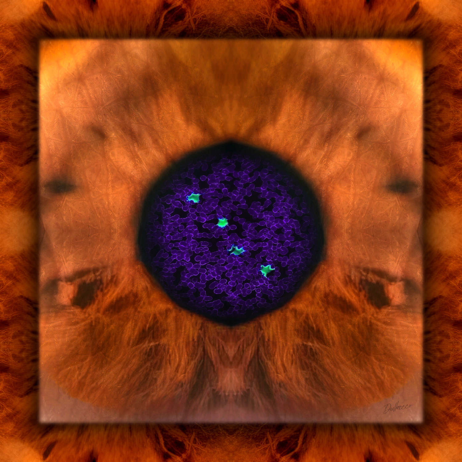
“UCR-DNA” is a BioArt series created as a tribute to former Vice Presidents for Research of the University of Costa Rica (1974-2025) through personalized cellular portraits. Each work emerges from the integration of cell imagery, bodily textures, and personal narratives obtained directly from the individuals portrayed, establishing a dialogue between biological identity, lived experience, and institutional legacy.
The project brings together cell biology, fluorescence microscopy, photomacrography, and digital visual composition to transform biological information into a poetic language of representation. Blood cells, iris textures, and skin microstructures become both artistic materials and symbolic traces of individuality, introducing a dimension of intimacy that cannot be artificially replicated. Through this process, the author explores the possibility of portraiture beyond physical appearance, seeking instead to evoke aspects of the subject’s essence, character, and contribution to the academic community.
Situated at the intersection of science, art, and memory, the series invites reflection on the relationship between biological uniqueness and personal history, proposing the human body as both archive and landscape. In doing so, this project transforms scientific observation into a commemorative act that celebrates the individuals whose work helped shape the development of research at the University of Costa Rica.
Cellular Portrait of Otto Rodrigo Gámez Lobo
Phlebotomy
Photomacrography
Fluorescence microscopy
Photomicrography
Digital mixed media BioArt
2025 - 25 x 25 cm
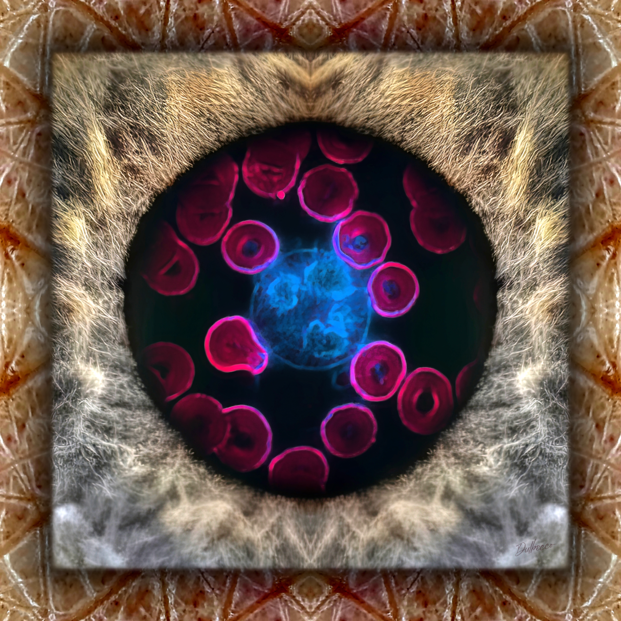
Description: The portrait was created using images obtained from the body of Mr. Luis Carlos Gámez Hernández, the son of Mr. Rodrigo. The periphery of the work displays the microscopic texture of the skin from his forearm, while the central panel reveals the macroscopic texture of his iris. Within the space of the pupil, a microscopic image of his erythrocytes (magenta) and one of his neutrophils is presented, with the latter’s genetic material appearing in blue after being labeled with a fluorescent intercalating agent that binds to the DNA double helix.
Author’s interpretation: The texture of Mr. Luis’s iris resembles the fur of a wolf, alluding to his father’s maternal surname. The central neutrophil and its DNA (blue) symbolize, respectively, Mr. Rodrigo and his profound passion for research, which he passed on to his colleagues, here represented by the surrounding erythrocytes imbued with that same DNA. This alludes to Mr. Rodrigo’s fundamental role in the consolidation of the Office of the Vice President for Research, extending a family legacy of public service established by his father, Mr. Uladislao, who served as Costa Rica’s Minister of Public Education on three occasions.
Cellular Portrait of Guillermo Chaverri Benavides
Phlebotomy
Photomacrography
Fluorescence microscopy
Photomicrography
Digital mixed media BioArt
2025 - 25 x 25 cm
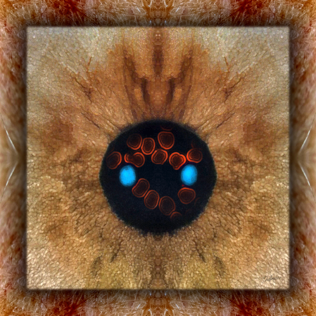
Description: The portrait was created using images obtained from the body of Mrs. Paquita Chaverri Jiménez, the daughter of Mr. Guillermo. The periphery of the work displays the microscopic texture of the skin from her forearm, while the central panel reveals the macroscopic texture of her iris. Within the space of the pupil, a microscopic image of her erythrocytes (red) and the cellular nuclei of two of her lymphocytes (light blue) is presented, which were labeled with a fluorescent intercalating agent that binds to the DNA double helix.
Author’s interpretation: Within the texture of Mrs. Paquita’s iris, an angel-like figure emerges, with two light-blue cellular nuclei at its center resembling eyes. This imagery alludes to the devotion and protection that Mr. Guillermo provided to his children and students. At the same time, these two lymphocytes represent the two renowned chemists bearing the Chaverri surname of that era: Mr. Guillermo and Mr. Gil.
Cellular Portrait of Primo Luis Chavarría Córdoba
Phlebotomy
Photomacrography
Fluorescence microscopy
Photomicrography
Digital mixed media BioArt
2025 - 25 x 25 cm
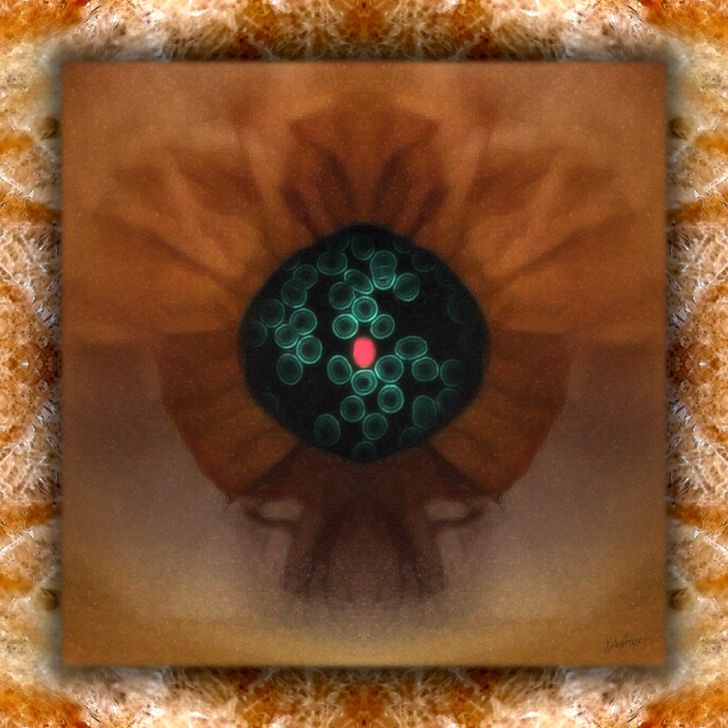
Description: The periphery of the work displays the microscopic texture of the skin from the subject’s forearm, while the central panel reveals the macroscopic texture of the iris. Within the space of the pupil appears a microscopic image of the subject’s erythrocytes (green) and the nucleus of one of his lymphocytes (salmon), labeled with a fluorescent intercalating agent that binds to the DNA double helix.
Author’s interpretation: Within the texture of the iris, a figure resembling a fruit emerges, containing the seed of knowledge, symbolized by Mr. Primo Luis’s DNA in salmon. This seed is surrounded by other green seeds, which represent the farmers he helped through his research in agronomy.
Cellular Portrait of Carlos Quesada Mateo
Phlebotomy
Photomacrography
Fluorescence microscopy
Photomicrography
Digital mixed media BioArt
2025 - 25 x 25 cm
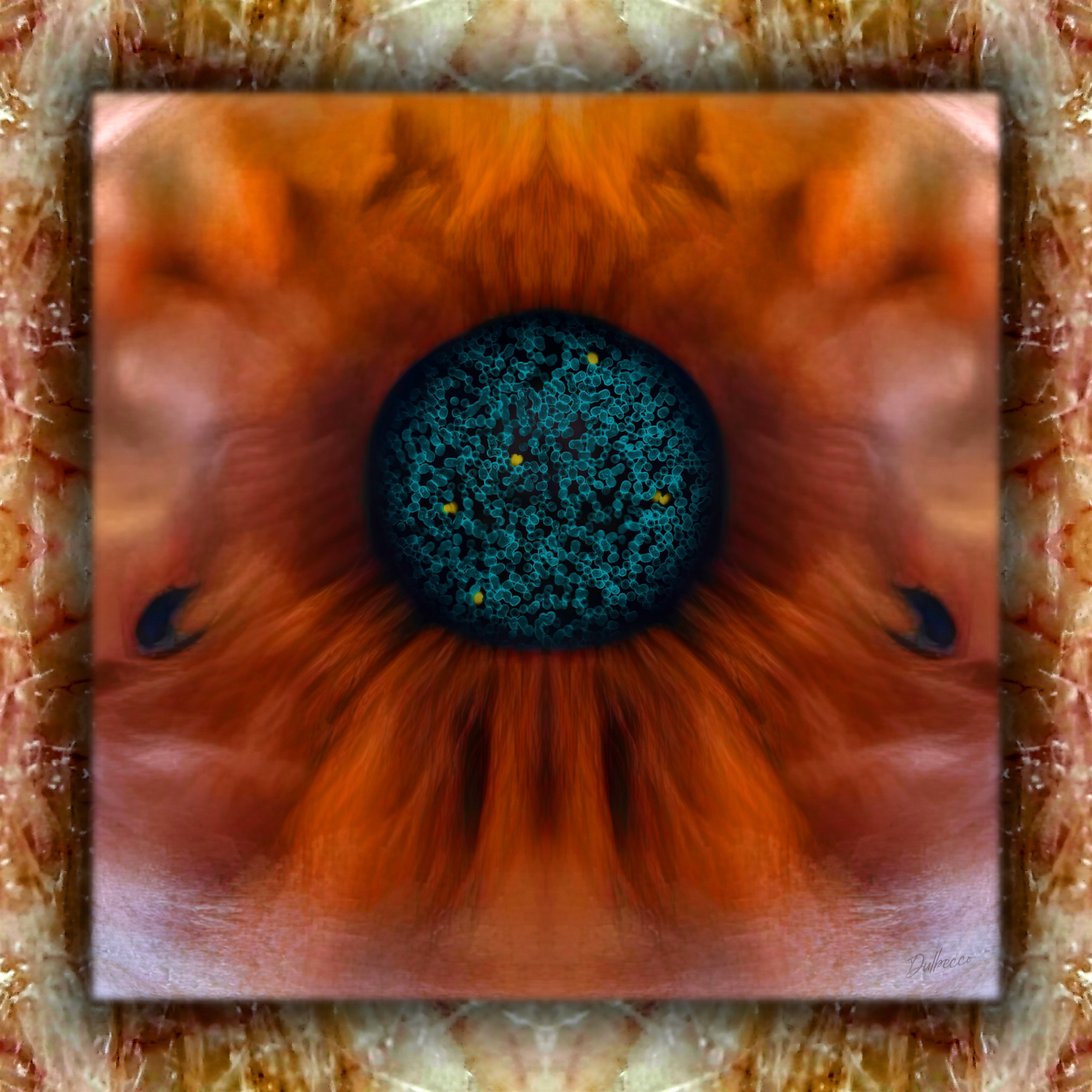
Description: The periphery of the work displays the microscopic texture of the skin from the subject’s forearm, while the central panel reveals the macroscopic texture of his iris. Within the space of the pupil, a microscopic image of the subject’s erythrocytes (turquoise) and the nuclei of five of his leukocytes (yellow) is presented. The latter were labeled with a fluorescent intercalating agent that binds to the DNA double helix.
Author’s interpretation: Within the texture of the iris, forms evoking a natural landscape with cascading waterfalls emerge, alluding to Mr. Carlos’s work on watershed management and sustainable development. The nuclei of his leukocytes are arranged as a network, symbolizing one of the defining achievements of his administration: the consolidation of the internet infrastructure at the University of Costa Rica.
Cellular Portrait of Yamileth González García
Phlebotomy
Photomacrography
Fluorescence microscopy
Photomicrography
Digital mixed media BioArt
2025 - 25 x 25 cm
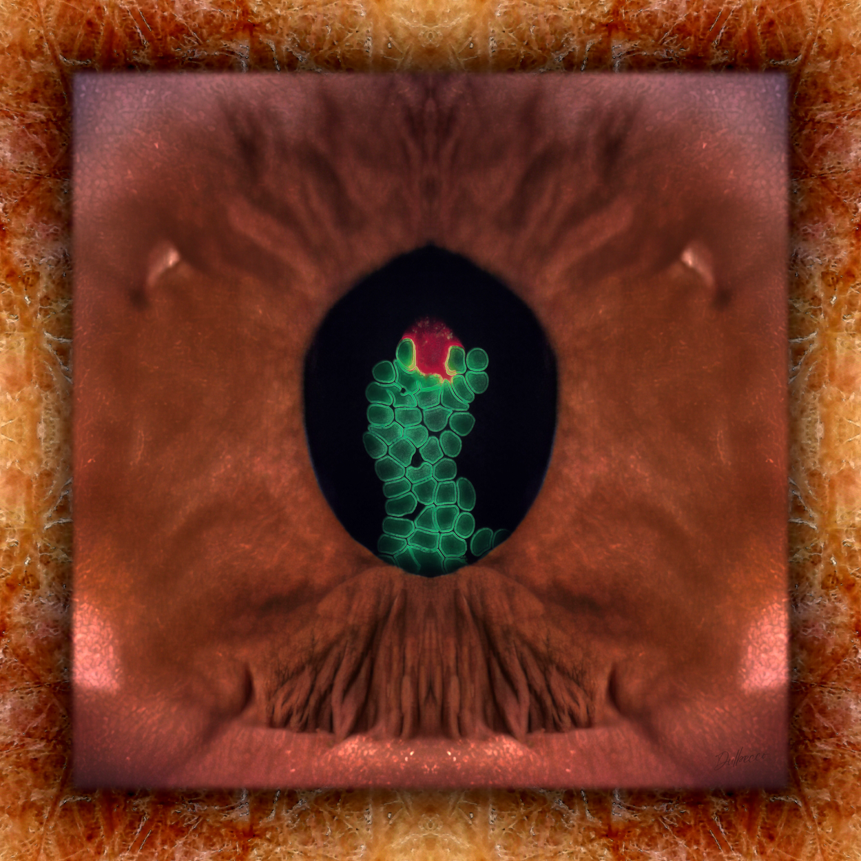
Description: The periphery of the work displays the microscopic texture of the skin from the subject’s forearm, while the central panel reveals the macroscopic texture of her iris. Within the space of the pupil appears a microscopic image of her erythrocytes (green) and one of her leukocytes, whose genetic material appears in red after being labeled with a fluorescent intercalating agent that binds to the DNA double helix.
Author’s interpretation: Within the texture of the iris, a dry landscape emerges, reminiscent of Orotina, Alajuela, the hometown of Mrs. Yamileth. The scene acquires a dreamlike quality, alluding to the aspirations of a young woman from a rural community who would go on to become the first woman to serve as Dean of the Graduate Studies System, Vice President for Research, and President of the University of Costa Rica. Along the same conceptual line, her blood cells resemble a pitahaya plant (Selenicereus undatus), a species characteristic of that environment. At its upper end appears the fruit in red (DNA), representing Mrs. Yamileth and one of her most distinctive qualities: mediating communication (yellow) among different actors within the university, here represented by the green erythrocytes.
Cellular Portrait of Henning Jensen Pennington
Phlebotomy
Photomacrography
Fluorescence microscopy
Photomicrography
Digital mixed media BioArt
2025 - 25 x 25 cm
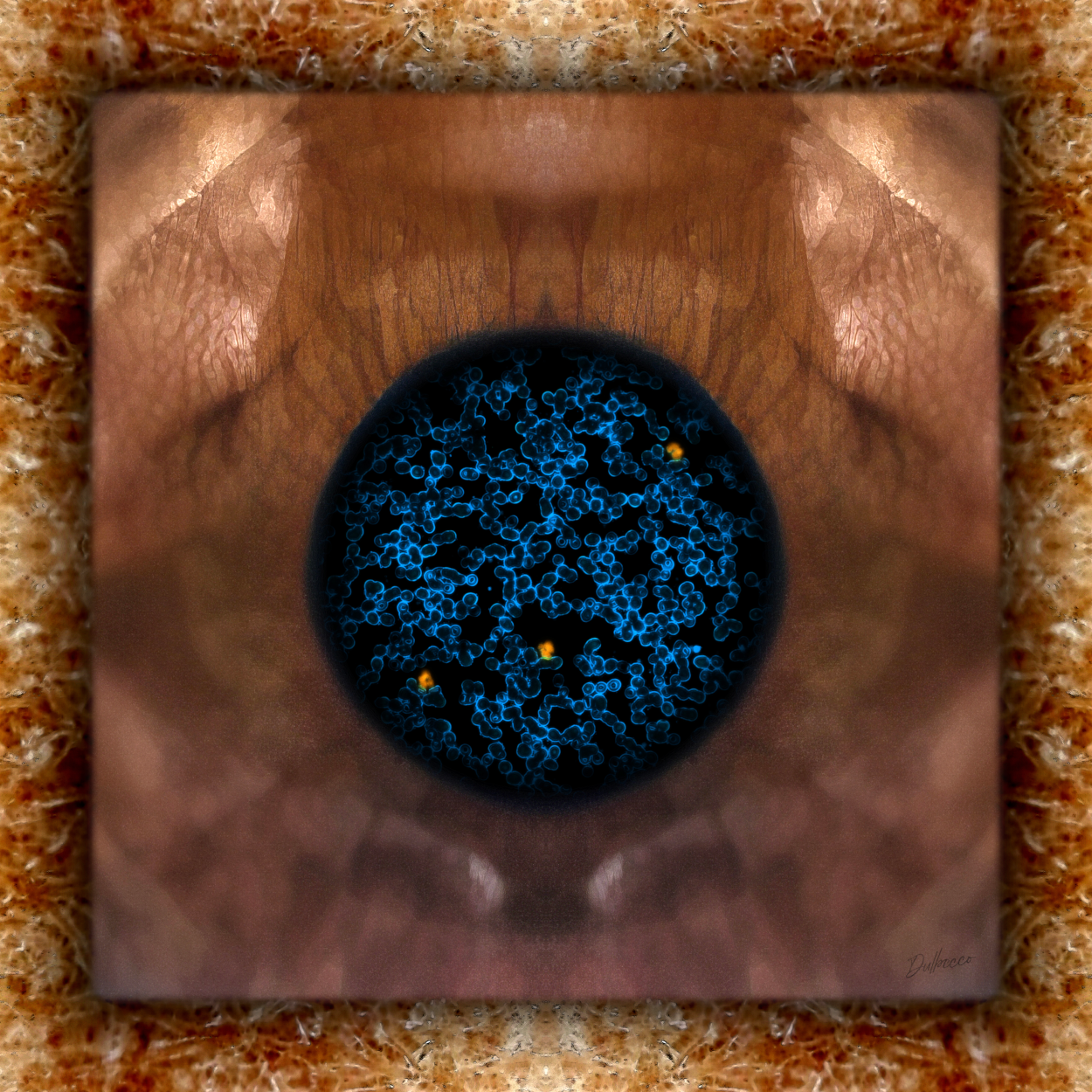
Description: The portrait was created using images obtained from the body of Mrs. Elena Jensen Villalobos, Mr. Henning’s daughter. The periphery of the work displays the microscopic texture of the skin from her forearm, while the central panel reveals the macroscopic texture of her iris. Within the space of the pupil, a microscopic image of her erythrocytes (blue) and three of her leukocytes is presented, with their nuclei appearing in orange after being labeled with a fluorescent intercalating agent that binds to the DNA double helix.
Author’s interpretation: Within Mrs. Elena’s iris texture, the duality between the conscious and the unconscious emerges, symbolized by the sharp upper portion and the blurred lower portion, respectively, alluding to her father’s profession as a psychologist. The arrangement of the orange cellular nuclei resembles the points of an upward-trending graph, representing the improvements in microbiology research achieved during Mr. Henning’s tenure as Vice President for Research and President of the University of Costa Rica.
Cellular Portrait of Ramiro Barrantes Mesén
Phlebotomy
Photomacrography
Fluorescence microscopy
Photomicrography
Digital mixed media BioArt
2025 - 25 x 25 cm
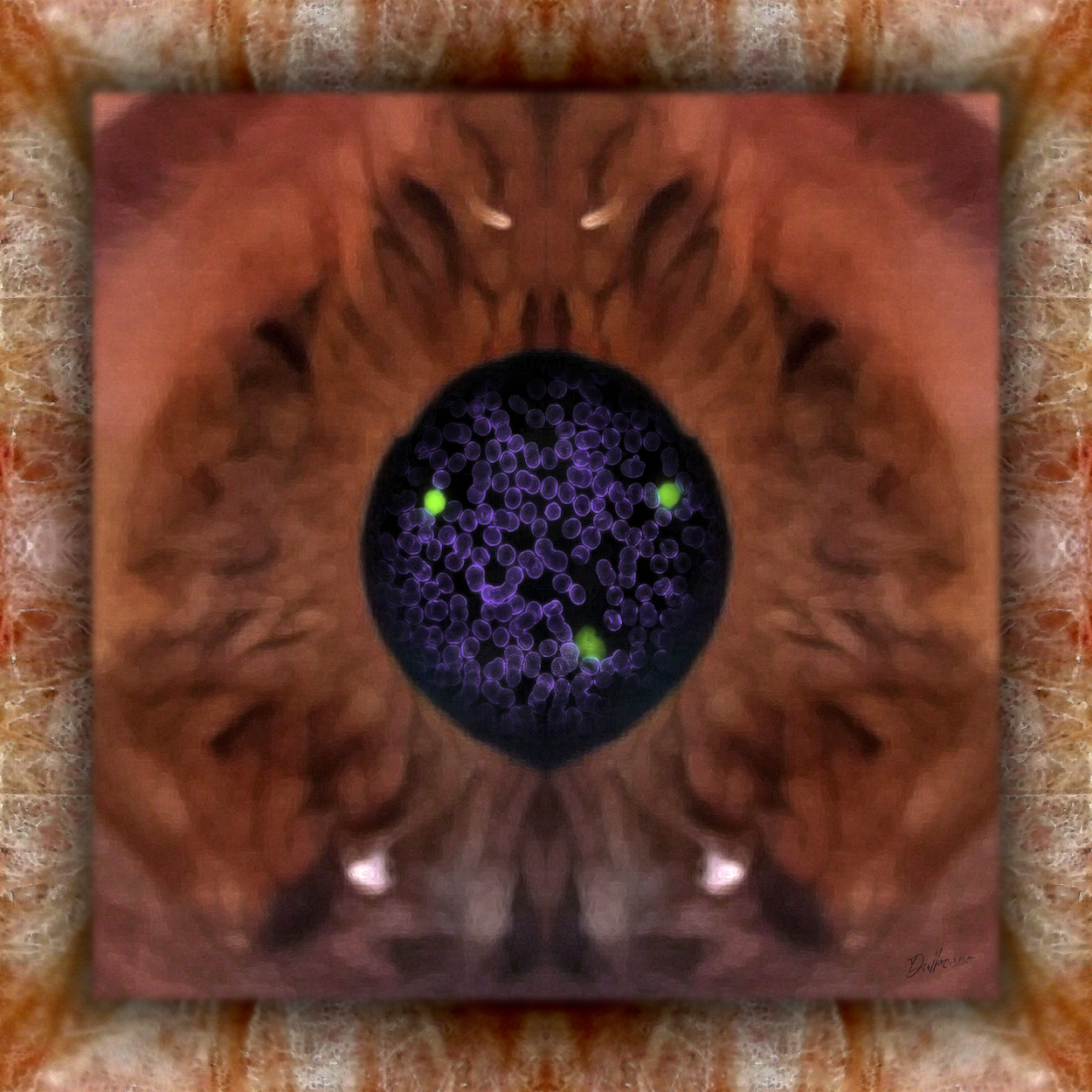
Description: The periphery of the work displays the microscopic texture of the skin from the subject’s forearm, while the central panel reveals the macroscopic texture of the iris. Within the space of the pupil, a microscopic image of the subject’s erythrocytes (purple) and the nuclei of his leukocytes (green) is presented. The latter were labeled with a fluorescent intercalating agent that binds to the DNA double helix.
Author’s interpretation: The texture of the iris forms an eye with reptilian features, alluding to Mr. Ramiro’s profession as a biologist. Within it emerges an entity that symbolizes his character and wisdom. The three green cellular nuclei represent his service as Vice President for Administration, Vice President for Academic Affairs, and Vice President for Research.
Cellular Portrait of Alice L. Pérez Sánchez
Phlebotomy
Photomacrography
Fluorescence microscopy
Photomicrography
Digital mixed media BioArt
2025 - 25 x 25 cm
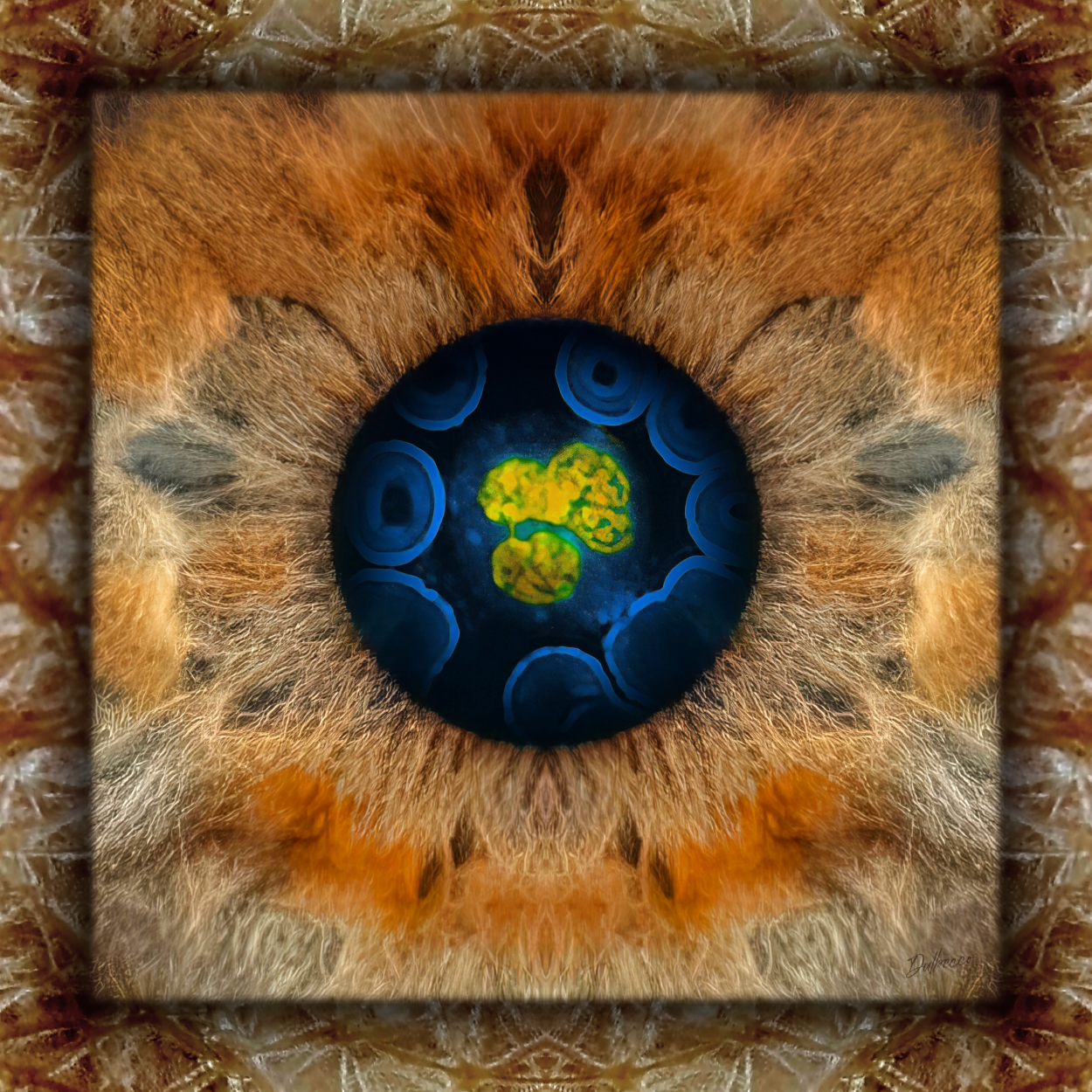
Description: The periphery of the work displays the microscopic texture of the skin from the subject’s forearm, while the central panel reveals the macroscopic texture of her iris. Within the space of the pupil, a microscopic image of her erythrocytes (blue) and one of her neutrophils is presented, with its nucleus appearing in yellow after being labeled with a fluorescent intercalating agent that binds to the DNA double helix.
Author’s interpretation: The subject’s iris displays a texture and coloration reminiscent of a Vancouver forest in autumn, representing the place where she completed her doctoral studies and her deep connection to the natural world through the research she conducts at the Center for Research in Natural Products. The yellow cellular nucleus appears to be undergoing metamorphosis, reflecting Mrs. Alice’s own nature as a scholar in constant transformation and growth.
Cellular Portrait of Fernando García Santamaría
Phlebotomy
Photomacrography
Fluorescence microscopy
Photomicrography
Digital mixed media BioArt
2025 - 25 x 25 cm
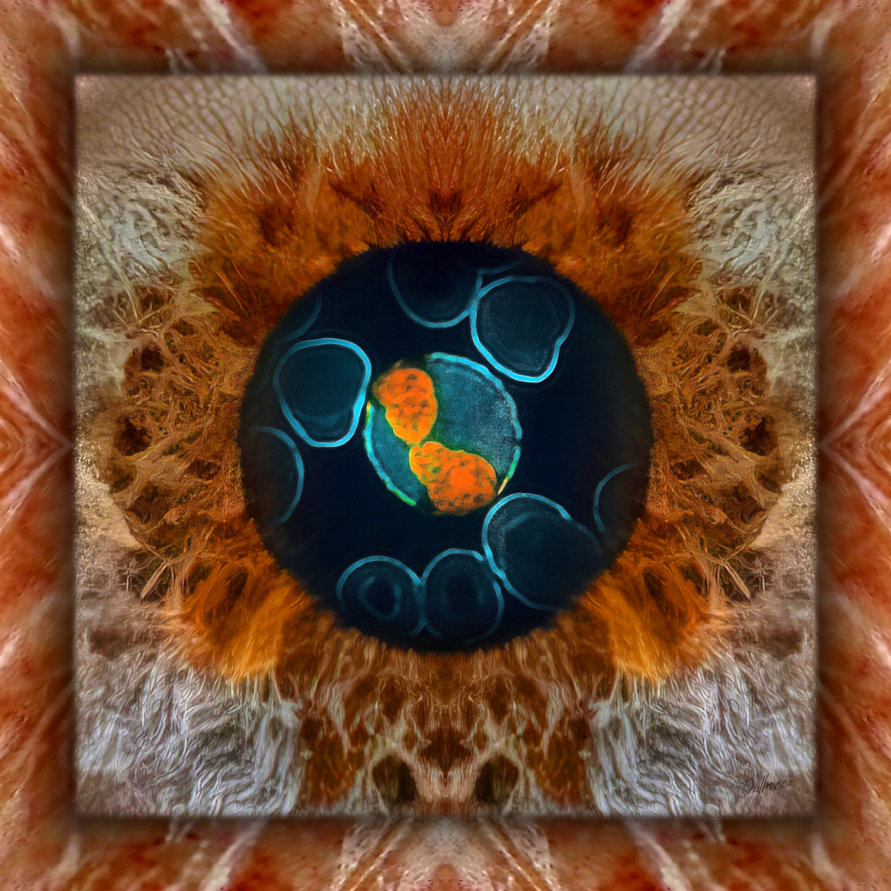
Description: The periphery of the work displays the microscopic texture of the skin from the subject’s forearm, while the central panel reveals the macroscopic texture of the iris. Within the space of the pupil, a microscopic image of the subject’s erythrocytes (turquoise) and one of his neutrophils is presented, with its nucleus appearing in orange after being labeled with a fluorescent intercalating agent that binds to the DNA double helix.
Author’s interpretation: Within the texture of the iris, a sunflower emerges, symbolizing Mr. Fernando’s dedication to the University of Costa Rica and his profound passion for research. The orange cellular nucleus displays two lobes that represent different stages of his life, alluding to his resilience and capacity for reinvention in the face of adversity.
Cellular Portrait of Adrián Pinto Tomás
Phlebotomy
Photomacrography
Fluorescence microscopy
Photomicrography
Digital mixed media BioArt
2025 - 25 x 25 cm
Description: The periphery of the work displays the macroscopic texture of the subject’s iris, while the central panel reveals a fusion of this texture with the microscopic structure of the skin from his forearm. Within the space of the pupil, a microscopic image of the subject’s erythrocytes (purple) and the nuclei of four of his leukocytes (cyan) is presented. The latter were labeled with a fluorescent intercalating agent that binds to the DNA double helix.
Author’s interpretation: The work is entirely related to Mr. Adrián’s research interests and greatest passion: the study of symbiotic interactions between microorganisms and social insects, such as ants. Within the textures of the subject’s iris and skin emerge forms reminiscent of macroscopic views of an ant’s body, while the DNA (cyan) visible in the image of his blood cells resembles the orderly movement of these insects as they travel in formation through their environment.
Cellular Portrait of María Laura Arias Echandi
Phlebotomy
Photomacrography
Fluorescence microscopy
Photomicrography
Digital mixed media BioArt
2025 - 25 x 25 cm
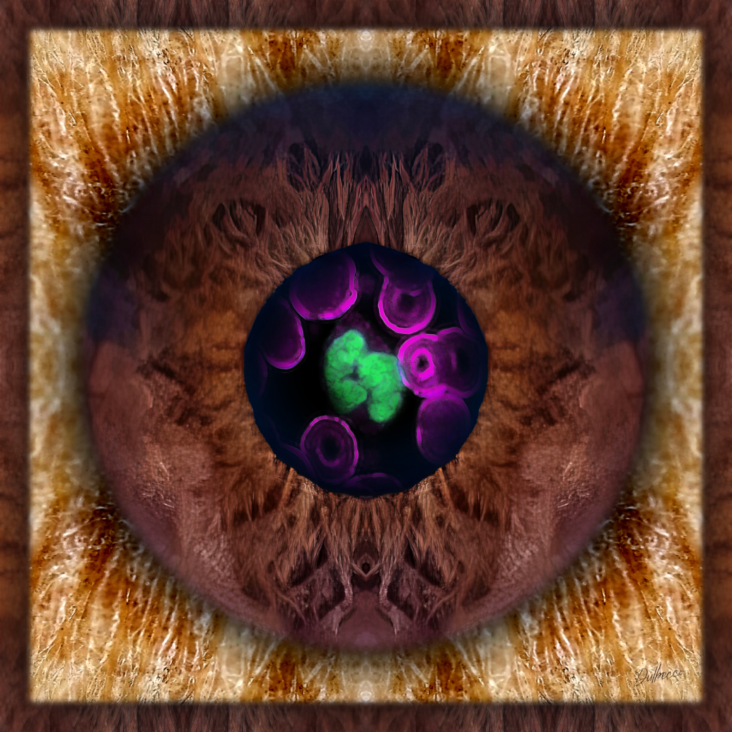
Description: The periphery and central circle of the work display the macroscopic texture of the subject’s iris, while the inner panel reveals the microscopic texture of the skin from her forearm. Within the space of the pupil, a microscopic image of her erythrocytes (magenta) and one of her neutrophils is presented, with its nucleus appearing in green after being labeled with a fluorescent intercalating agent that binds to the DNA double helix.
Author’s interpretation: The subject’s iris displays a wood-like texture that symbolizes her strength and character, followed by the microscopic detail of her skin, which, like a delicate veil, evokes her kindness and collaborative nature. The green cellular nucleus exhibits two lobes representing the two cerebral hemispheres, alluding to her multifaceted nature and the diversity of roles she has fulfilled throughout her life as a mother, scholar, entrepreneur, and figure within university politics.
Cellular Portrait of José Moncada Jiménez
Phlebotomy
Photomacrography
Fluorescence microscopy
Photomicrography
Digital mixed media BioArt
2025 - 25 x 25 cm
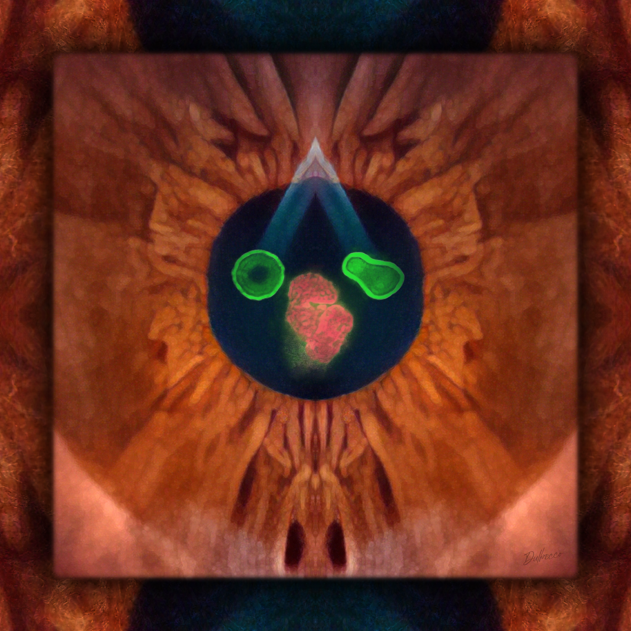
Description: The periphery of the work displays a fusion of the microscopic structure of the skin from the subject’s forearm with the macroscopic texture of the iris of one of his eyes, while the central panel reveals the latter texture in isolation. Within the space of the pupil, a microscopic image of his erythrocytes (green) and the nucleus of one of his neutrophils (salmon) is presented. The latter was labeled with a fluorescent intercalating agent that binds to the DNA double helix.
Phlebotomy: Francisco Vega Aguilar / Music composition & performance: Alonso Saavedra Coles / Photomacrography & video production: Gustavo López Marín
Author’s interpretation: The work portrays the critical period currently faced by the University of Costa Rica in response to pressures from the national goverment, a situation represented by the dark abyss visible in the lateral panel. The central panel represents Mr. José, who, through his position, must serve as a bridge to ensure that research activities continue amid the crisis. The nucleus of the central neutrophil resembles a fetus, symbolizing the Office of the Vice President for Research, while the green erythrocytes, displaying different morphologies, represent both the traditional approach to research (round), which has yielded results to date, and the innovative approach (irregular), which must be embraced to ensure the continuation of high-quality research under current circumstances.