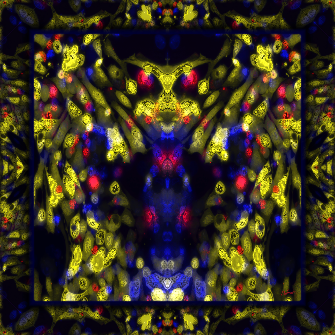
“Cellular Entities” is an ongoing BioArt series based on a particular vision of the natural world, where the application of bilateral symmetry to microscopic cellular beings invites the human brain to perceive visual analogies with patterns and forms of the macroscopic world.
The author inserted new genes into animal cells to enable recombinant color expression through fluorescent proteins and then collaborated with these genetically modified organisms to create an in vitro microscopic environment, which he explored using live-cell fluorescence microscopy. The resulting images (base photomicrographs) were used as inputs for a visual programming network developed by the author and employed during the same compositional process to create the main artworks and animate the static forms of the original images. Finally, dedicated music was created for each work (audiovisual BioArt). To avoid introducing biases arising from the artist’s own interpretation, the main works were assigned simple numerical designations. Additionally, some pieces are accompanied by derivative artworks portraying particular cellular entities that revealed themselves to the author during his own contemplative process.
The project is based on a psychological phenomenon known as pareidolia, which consists of the recognition of meaningful patterns, such as faces and bodies, within random visual information. In this way, it draws upon what is known in visual semiotics as mnemonic attractors: collections of mental images accumulated within an individual’s associative memory throughout life experiences that enable the retention and recollection of specific memories. This completely personalizes the interpretation of each work, making it dependent upon the genetic, cultural, and ontogenetic background of the viewer. It is here that the project proposes a new direction within BioArt, one that not only employs biological elements in its technique and discourse, but also incorporates the social and epigenetic dimensions of the viewer into the artistic process.
This proposal invites people to pause, interpret, and reflect on each work, amidst a modernity characterized by the constant overstimulation of the senses through screens. In this way, the contemplative system becomes the antithesis of a disposable consumption model, opening a window to abstraction and a reconnection with the natural world, which for many has been relegated to the background due to technology. Each work represents a portal through which different entities from the viewer’s subconscious can manifest.
Cellular Entities I
Cell culture
Genetic engineering
Fluorescent proteins
Live-cell fluorescence imaging
Digital mixed media BioArt
2023 - 50 x 50 cm
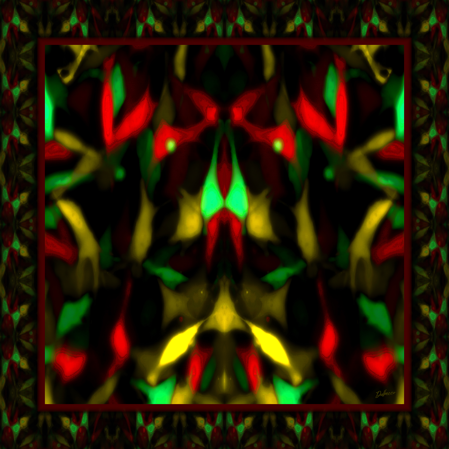
Base photomicrograph – 18 x 25 cm

Audiovisual BioArt - 4K
Description: Fluorescence imaging of baby hamster kidney cells genetically modified to express different fluorescent proteins (green, red, and yellow).
Cellular Entities II
Cell culture
Genetic engineering
Fluorescent proteins
Live-cell fluorescence imaging
Digital mixed media BioArt
2023 - 40 x 40 cm
Base photomicrograph – 15 x 20 cm
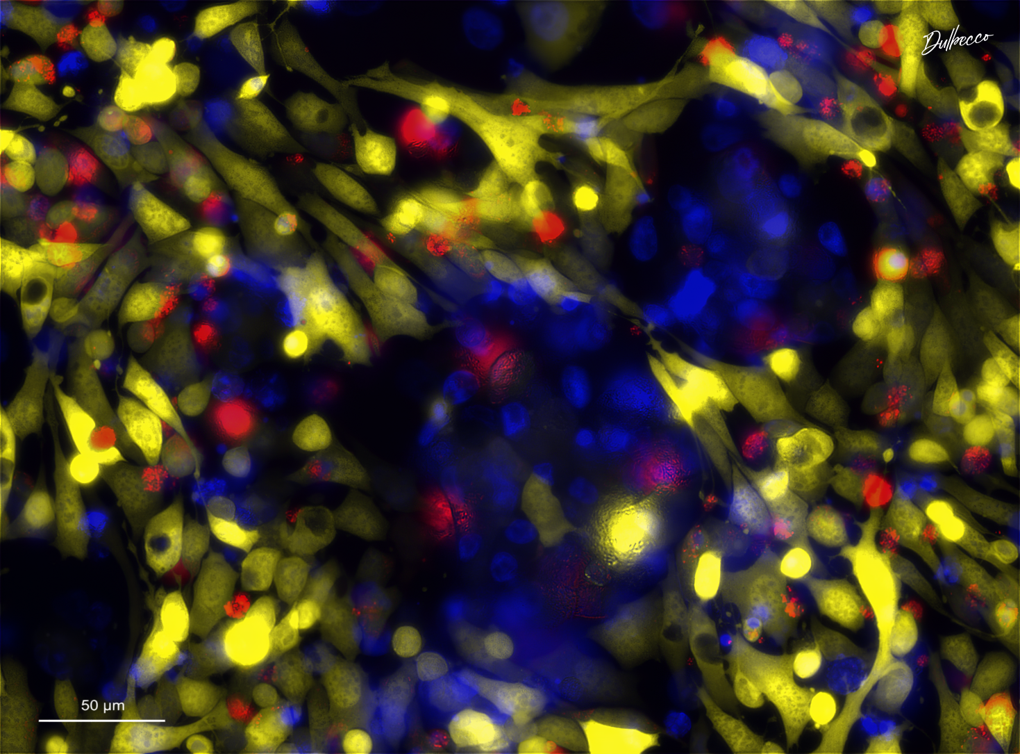
Audiovisual BioArt - 4K
Description: Fluorescence imaging of a co-culture of human embryonic kidney cells genetically modified to express a fluorescent protein (yellow) with human cervical cancer cells. DNA intercalating agents were used to label nuclei (blue) and cell death (red).
Cellular Entities III
Cell culture
Genetic engineering
Fluorescent proteins
Live-cell fluorescence imaging
Digital mixed media BioArt
2023 - 50 x 50 cm

Base photomicrograph – 18 x 25 cm

Audiovisual BioArt - 4K
Description: Fluorescence imaging of human embryonic kidney cells genetically modified to express fluorescent proteins (white and cyan). A specific fluorescent probe was used to label mitochondria (magenta).
Cellular Entities IV
Cell culture
Genetic engineering
Fluorescent proteins
Live-cell fluorescence imaging
Digital mixed media BioArt
2023 - 50 x 50 cm

Base photomicrograph – 18 x 25 cm

Audiovisual BioArt - 4K
Description: Fluorescence imaging of baby hamster kidney cells genetically modified to express fluorescent proteins (yellow, red, and green). A DNA intercalating agent was used to label cell death (white)
Cellular Entities V
Cell culture
Genetic engineering
Fluorescent proteins
Live-cell fluorescence imaging
Digital mixed media BioArt
2023 - 50 x 50 cm

Base photomicrograph – 18 x 25 cm

Audiovisual BioArt - 4K
Description: Fluorescence imaging of baby hamster kidney cells genetically modified to express fluorescent proteins (pink and white). DNA intercalating agents were used to label nuclei (green) and cell death (yellow).
Cellular Entities VI
Cell culture
Genetic engineering
Fluorescent proteins
Live-cell fluorescence imaging
Digital mixed media BioArt
2024 - 40 x 40 cm

Base photomicrograph – 14 x 19 cm

Audiovisual BioArt - 4K
Description: Fluorescence imaging of human embryonic kidney cells genetically modified to express a fluorescent yellow protein. Specific fluorescent probes were used to label nuclei (green) and mitochondria (magenta).