
“The Scientist” is an ongoing BioArt series currently composed of twenty-three fluorescence photomicrographs captured by the author over eighteen years of active scientific research involving animal cells and viruses. The works are micropoetics: abstractions, analogies, and allegories of the macroscopic world within the microbiological universe, and vice versa. The project also reflects the author’s contemplations and inner experiences arising from his sustained dialogue with the microscopic entities with which he works.
This gallery merges science and art by repurposing cutting-edge techniques from cellular and molecular biology to address contemporary issues, including climate change, environmental pollution, pandemics, virtuality, identity, human rights, and armed conflicts, among others, through a biopoetic and conceptual lens. All methodologies and protocols used to create the works were developed by the author in a handcrafted manner and have been published in his scientific articles. As such, the gallery also reflects the author’s evolution as a scientist, both in terms of his biotechnological expertise and the progressive advancement of the laboratory instrumentation supporting his research at the University of Costa Rica.
Tchaikovsky Op.20
Cell culture
Immunofluorescence
Digital photomicrography
2008 - 30 x 38 cm

Description: Fluorescence microscopy of human umbilical artery smooth muscle cells infected with Dengue Virus (green). Cytoplasms of non-infected cells are shown in red and nuclei in blue.
Author’s interpretation: Microscopic allegory of Tchaikovsky’s famous ballet “Swan Lake”. The Black Swan is embodied by the infected cell (green), which emerges onto the stage surrounded by the other dancers.
B-612
3D cell culture
Immunohistochemistry
Digital photomicrography
2008 - 30 x 38 cm

Description: Fluorescence microscopy of a 3D culture of human umbilical vein endothelial cells and human umbilical artery smooth muscle cells (red). The cell nuclei are shown in blue.
Author’s interpretation: When I return to the places of my childhood after many years, I find that the world that once felt vast and magical has, through the altered perspective of adulthood, become unexpectedly small and real.
Kanagawa
Cell culture
Fluorescence microscopy
Digital photomicrography
2009 - 30 x 38 cm

Description: Fluorescence microscopy of human carcinoma cells showing cytoplasms in cyan and nuclei in magenta.
Author’s interpretation: Microscopic allegory of the famous woodblock print from the 19th century “The Great Wave off Kanagawa” by Katsushika Hokusai. The piece also represents the rise in the levels of the sea due to the climate change that is directly affecting many islands in the Pacific Ocean.
Territory
Cell culture
Immunofluorescence
Digital photomicrography
2010 - 30 x 38 cm

Description: Fluorescence microscopy of a co-culture of human umbilical vein endothelial cells (orange) with human umbilical artery smooth muscle cells (green). The cell nuclei are shown in blue.
Author’s interpretation: These cell lineages normally coexist and collaborate within the walls of blood vessels. However, when removed from their native tissue and placed together in a culture dish in vitro, they begin to compete for space and resources. Are our behaviors as human beings a reflection of processes rooted at the cellular level? This work is a microscopic abstraction of the Israeli-Palestinian conflict.
Seahorse
Cell culture
Immunofluorescence
Digital photomicrography
2011 - 30 x 38 cm

Description: Fluorescence microscopy of monkey kidney epithelial cells infected with Herpes Simplex Virus 1 (green).
Author’s interpretation: Microscopic allegory of a male seahorse that represents all the marine wildlife in danger by the warming of the oceans due to climate change.
Reef
Cell culture
Immunofluorescence
Digital photomicrography
2012 - 30 x 38 cm

Description: Fluorescence microscopy of human carcinoma cells infected with Human Parainfluenza Virus (green). Cytoplasms of non-infected cells are shown in red and nuclei in blue.
Author’s interpretation: Coral reefs are important ecosystems that enable life underwater and provide a crucial source of food and jobs for millions of people. They also protect coastlines from storms and erosion by reducing the power of waves hitting the coast. Climate change is dramatically affecting coral reefs all over the world. Its conservation and restoration must be one of the primary goals for present and future generations.
Climate Change
Cell culture
Immunofluorescence
Digital photomicrography
2013 - 30 x 38 cm

Description: Fluorescence microscopy of Dengue Virus plaques on monkey kidney epithelial cells (green). Cytoplasms of non-infected cells are shown in red and nuclei in blue.
Author’s interpretation: Microscopic allegory of the environmental problem we are facing in the present. Our world is represented in a single well with cells that suffer the drastic changes in the culture conditions occasioned by a viral infection (green), that spreads over this microscopic landscape like twisters and hurricanes do in the affected areas of our planet.
The Chernobyl Fungi
Cell culture
Immunofluorescence
Digital photomicrography
Digital BioArt
2014 - 30 x 38 cm

Description: Fluorescence microscopy of monkey kidney epithelial cells infected with Herpes Simplex Virus 1 (green). Cytoplasms of non-infected cells are shown in red and nuclei in blue.
Author’s interpretation: Microscopic allegory of the radiotropic fungi discovered in the radioactive exclusion zone surrounding the Chernobyl nuclear power plant. The work reflects on the paradoxical creativity of life: how human errors can open new evolutionary pathways for other species, and how organisms that emerge from our catastrophes may ultimately help bioremediate what we have damaged.
Ephemeral
Cell culture
Fluorescence microscopy
Digital photomicrography
2015 - 30 x 38 cm

Description: Fluorescence microscopy of human umbilical vein endothelial cells showing cytoplasms in purple and nuclei in magenta.
Author’s interpretation: Contemporary consumer society always has something new to display, encouraging us to forget what has just passed. In the world of the attention economy, we seem to inhabit a perpetual present, unable to fully absorb the incessant stream of information flowing across our screens. In this context, the cellular forms within the artwork evoke the transitory nature of contemporary ideas, whose existence appears to fade with the same speed and volatility as a dispersing gas.
Pollock
Cell culture
Immunofluorescence
Digital photomicrography
Digital BioArt
2016 - 30 x 38 cm

Description: Fluorescence microscopy of rhesus monkey cells (magenta) infected with Zika Virus (yellow). Cell nuclei are shown in cyan.
Author’s interpretation: Microscopic abstraction of the drip technique, a splashing liquid paint method used by American artist Jackson Pollock, a key figure of the Abstract Expressionist movement in the second half of the 20th century. Cell textures from different microscopic fields were overlaid on a digital canvas.
1P/Halley
Cell culture
Immunofluorescence
Digital photomicrography
2016 - 30 x 38 cm

Description: Fluorescence microscopy of monkey kidney epithelial cells infected with Herpes Simplex Virus 1 (white).
Author’s interpretation: Microscopic allegory of comet 1P/Halley. Edmond Halley (1656-1742) stole a victory from superstition and contributed new knowledge to science by mathematically predicting the period of his eponymous comet.
Lucem Aspicio
Cell culture
Immunofluorescence
Digital photomicrography
2017 - 48 x 60 cm

Description: Fluorescence microscopy of human umbilical artery smooth muscle cells (cyan) infected with Dengue Virus (yellow). Cell nuclei are shown in magenta.
Author’s interpretation: Microscopic allegory of the University of Costa Rica. State institutions of higher education in Costa Rica not only contribute to the country’s development through the training of highly skilled professionals. Public universities also serve as centers for knowledge generation and patronage for thinkers, scientists, and artists who, through their work, create portals to new horizons and realities.
The Spectrum
Cell culture
Genetic engineering
Live-cell fluorescence imaging
Digital photomicrography
2018 - 30 x 41 cm

Description: Fluorescence imaging of baby hamster kidney cells genetically modified to express different fluorescent proteins (green, red, and yellow). Cell nuclei were labeled with a DNA intercalating agent (blue).
Author’s interpretation: Microscopic abstraction of the spectrum of identities and personalities of biological entities. We are not static organisms, but rather homeostatic ones in constant evolution.
Tropical Spring
Cell culture
Genetic engineering
Live-cell fluorescence imaging
Digital photomicrography
2019 - 30 x 40 cm

Description: Viral plaque assay in monkey kidney cells infected with a fluorescent version of Herpes Simplex Virus 1 (violet). DNA intercalating agents were used to label chromatin condensation (cyan) and cell death (white), respectively.
Author’s interpretation: Microscopic allegory of the ‘Pink poui’ (Tabebuia rosea), which blooms during the summer in Costa Rica and beautifies rural and urban landscapes with its hues.
EVE
Cell culture
Immunofluorescence
Digital photomicrography
Social BioArt
2020 - 30 x 38 cm

Description: Fluorescence microscopy of a co-culture of human umbilical vein endothelial cells (orange) with human umbilical artery smooth muscle cells (green).
Author’s interpretation: Homage to African culture and to Afro-descendant people, the mother of humanity. Inadvertently spread throughout the world and in constant struggle against the systematic discrimination that has existed for centuries. The artwork was revealed to the author during a time of constant tension and protest by human rights movements.
Virtual Meeting
Cell culture
Immunofluorescence
Digital photomicrography
Digital BioArt
2021 - 30 x 38 cm

Description: Fluorescence microscopy of human nerve cells infected with Herpes Simplex Virus 1.
Author’s interpretation: Microscopic allegory of our modern meetings. Neurons from different microscopic fields were placed together by digital means in a virtually created microscopic space.
Collective Illusion
Cell culture
Genetic engineering
Live-cell fluorescence imaging
Digital photomicrography
2021 - 40 x 58 cm

Description: Fluorescence-based SARS-CoV-2 neutralization assay. Shown are monkey kidney cells genetically modified to express both a fluorescent protein (orange) and the ACE2 receptor used by SARS-CoV-2 to enter the cell. Cell nuclei were labeled with a fluorescent intercalating agent (blue), and live-cell fluorescence images were acquired 72 hours after the addition of a lentiviral vector carrying the SARS-CoV-2 Spike protein on its envelope. The vector had been previously incubated with serum from a patient who had received a COVID-19 vaccine.The vector induces the expression of a green fluorescent protein that should not be produced if the patient possessed neutralizing antibodies against SARS-CoV-2 capable of blocking the Spike protein and, consequently, preventing the vector from entering the cell. Therefore, the green fluorescence observed in the image indicates that the patient lacked neutralizing antibodies against SARS-CoV-2 despite having received a COVID-19 vaccine.Finally, 49 independent fluorescence images were acquired at a total magnification of 200X and assembled into a single high-definition image.
Author’s interpretation: This work highlights how the mere act of receiving a vaccine does not guarantee protection from an immunological standpoint, as many patients fail to produce high levels of neutralizing antibodies, as demonstrated by the neutralization assay presented. In these cases, vaccination becomes a kind of conceptual placebo with more political and economic purposes than public health benefits.
The Whole
Cell culture
Genetic engineering
Live-cell fluorescence imaging
Digital photomicrography
2022 - 30 x 41 cm

Description: Fluorescence imaging of genetically modified human cervical carcinoma cells to express a fluorescent protein (white). The nuclei, mitochondria, and cytoskeleton were stained with fluorescent probes (green, red, and purple, respectively). These cells originally were part of the tumor that took the life of Henrietta Lacks (1920-1951). The artwork is dedicated to her memory.
Author’s interpretation: From my perspective, the cells of a deceased individual that continue to divide under laboratory conditions offer biological evidence of their autonomy as living entities. I often wonder whether our cells have any notion that they belong to us. Perhaps they do not. Perhaps, in the same way, we remain unaware that we ourselves belong to something greater within a vast fractal continuum, in which every level of existence is both an individual and a part of a larger whole…
Bolivar
Cell culture
Genetic engineering
Live-cell fluorescence imaging
Digital photomicrography
Social BioArt
2022 - 28 x 40 cm

Description: Fluorescence imaging of a mixed culture of genetically modified human embryonic kidney cells expressing a yellow fluorescent protein, together with human cervical cancer cells. DNA-intercalating agents were used to stain the nuclei (blue) and dead cells (red).
Author’s interpretation: Microscopic allegory of the political instability experienced in Colombia, Venezuela, and Ukraine in 2022. The artwork symbolizes the struggle of three peoples moving forward in pursuit of freedom. The human carcinoma cells, stained blue, represent oppressive political systems. The innocent populations suffering under these regimes are symbolized by the human embryonic kidney cells in yellow. The dead cells, stained red, allude to the martyrs of these injustices.
Lo Fatal
Cell culture
Immunofluorescence
Digital photomicrography
Social BioArt
2023 - 38 x 30 cm

Description: Fluorescence microscopy of human umbilical artery smooth muscle cells (red) infected with Dengue Virus (green). Cell nuclei are shown in purple.
Author’s interpretation: This work establishes an analogy between the political system currently governing the Republic of Nicaragua and a lethal viral infection within a cellular system. The map of the country is microscopically represented through a photomicrograph of human cells infected with Dengue virus and stained using an immunofluorescence technique. Its title derives from Rubén Darío’s poem of the same name, in which the uncertainty of life is explored from the perspective of a conscious being. A second analogy is drawn with Nicaraguan youth, who grow up within a repressive system where those in power transform mere subjectivities into absolute and irrefutable truths.
Superior Order
Cell culture
Genetic engineering
Live-cell fluorescence imaging
Digital photomicrography
2024 - 30 x 41 cm
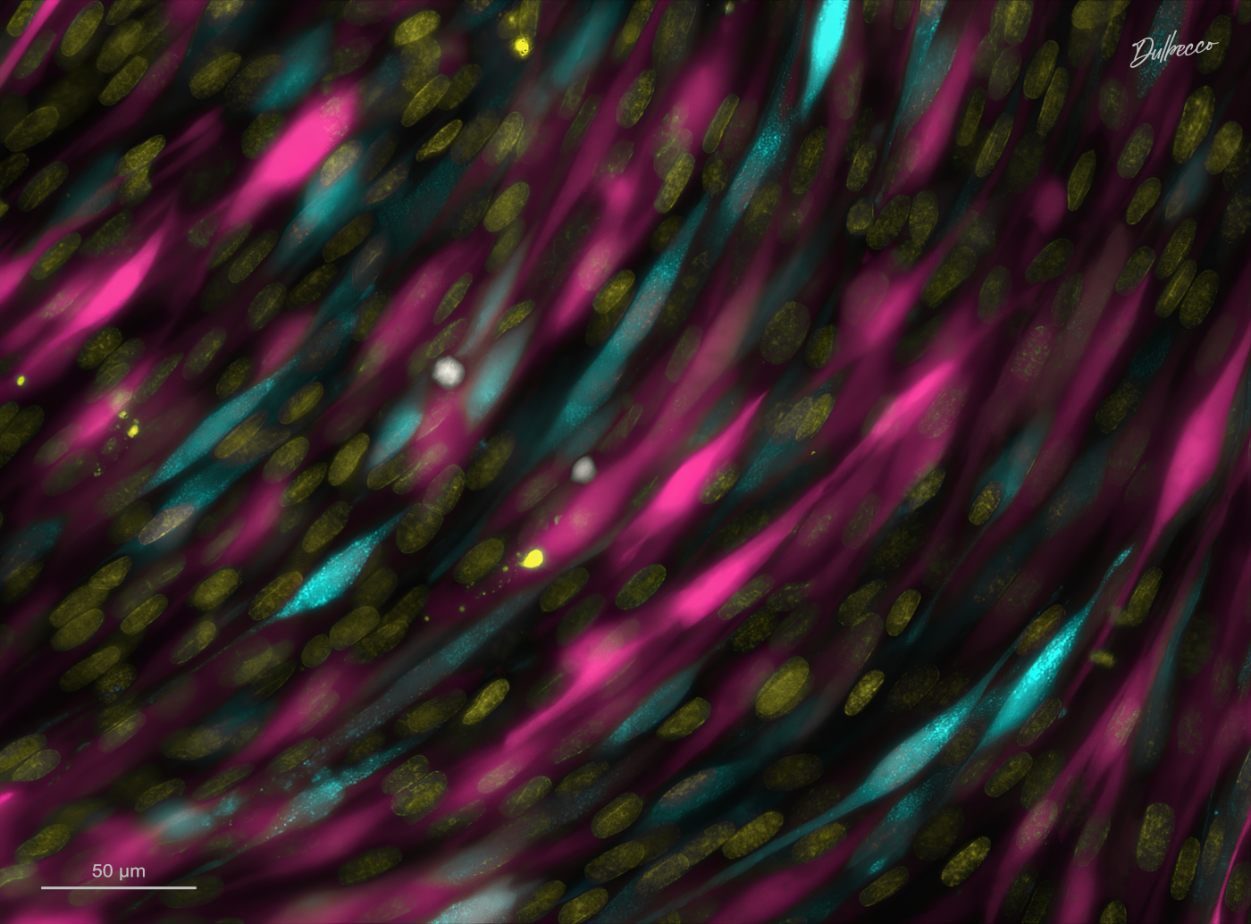
Description: Fluorescence imaging of genetically modified hamster kidney fibroblasts expressing cytoplasmic cyan and magenta fluorescent proteins. Cell nuclei are shown in yellow.
Author’s interpretation: Initially, cells wander through space as autonomous beings, tracing seemingly random paths. Yet as they multiply, an unseen order begins to emerge. Individual trajectories converge into a shared architecture, and what once appeared as a scattered gathering gradually unfolds into a living tissue. This microscopic landscape suggests that living entities, no matter how independent, may ultimately be part of a larger whole: a quiet fractal of existence unfolding from the collective.
Who Are You?
Cell culture
Genetic engineering
Live-cell fluorescence imaging
Digital photomicrography
2025 - 59 x 45 cm
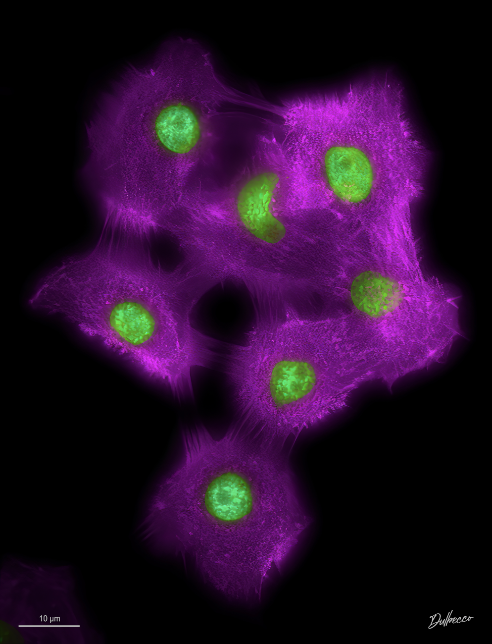
Description: Fluorescence imaging of human carcinoma cells expressing genetically encoded fluorescent sensors for the cytoskeleton (purple) and nuclei (green).
Author’s interpretation: Do you really know who you are? We believe we know ourselves because we inhabit our bodies, but not everything that passes through our minds makes sense. We constantly create fictions and false interpretations. We must learn to filter our thoughts. After all, there is no self…
Blackout
Cell culture
Live-cell fluorescence imaging
Digital photomicrography
Social BioArt
2026 - 30 x 41 cm
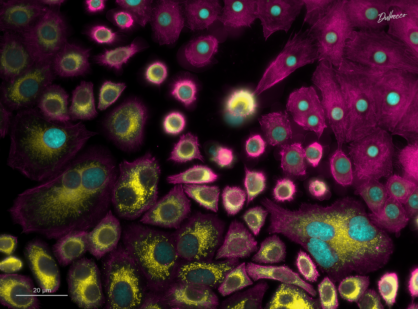
Description: Fluorescence imaging of human cervical carcinoma cells labeled with fluorescent probes for the cytoskeleton (magenta), nucleus (cyan), and mitochondria (yellow).
Author’s interpretation: Microscopic allegory of contemporary parliaments shaped by rigid partisanship, where individual legislative freedom is subordinated to strict party discipline and entrenched ideological blocs. The image depicts two cellular lineages standing divided. One, “lit,” occupies the lower region of the image, displaying large nuclei and abundant mitochondria. The other, “darkened,” resides in the upper region, marked by small nuclei and the mysterious absence of mitochondrial labeling. This population of “darkened” cells seeks to recruit new adherents to its cause among the cells of mixed morphology that linger along the border between both lineages.